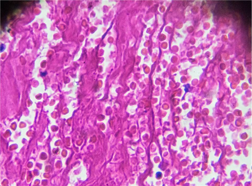
The first step after creating a disease model and collecting tissue samples is histological analysis. This approach includes anything from broad behavioral observations to in-depth research into the disease, cell biology, and molecular biology. The outcomes of your challenging front-end investigations are guaranteed by a thorough and stable histological examination, which also lays a solid foundation for the success of your later studies.
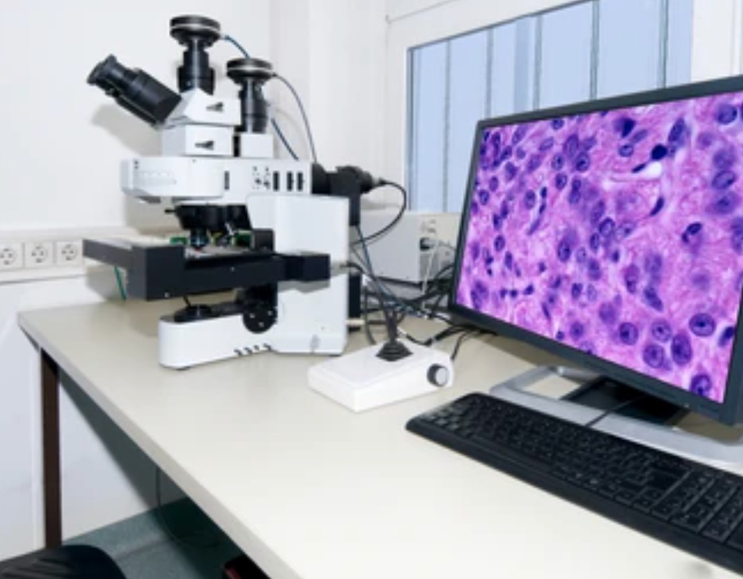
The use of computers and networks in the discipline of pathology is referred to as digital pathology. It uses a fully automated microscope or optical magnification system to scan and acquire high-resolution digital images, and then a computer is used to automatically stitch and process the images in multiple fields of view with high precision to obtain high-quality visual data for use in all areas of pathology.
Our company provides customers with professional high-throughput histology and digital pathology analysis services for qualitative and quantitative assessment of drug efficacy.
Service Overview

Our company currently provides a comprehensive spectrum of histology and digital pathology services from sample preparation to detailed and trustworthy research results for our clients globally thanks to our considerable laboratory experience and cutting-edge theoretical background. We use automated immunohistochemistry, immunofluorescent labeling, and digital analysis to quickly shed light on the mechanisms underlying medication action as well as its efficacy.
To guarantee that the outcomes of our meticulous validation and quality control methods are correct and consistent, we classify tissues, identify subcellular structures, and analyze geographic distribution and closeness using industry standards.
Our company's rigorously validated histology platform provides a comprehensive range of histological features that enable you to:
- Correctly display the spatial distribution and localization of specific cellular components in the appropriate histological context.
- Assess drug efficacy by detecting the up- or down-regulation of drug activity and disease markers in target tissues and other sites.
- Accurately localize targets in tumor tissues.
- Increase the likelihood of preclinical and clinical success of your drug.
Customers typically supply glass microscope slides for scanning and analysis or preserved or frozen tissue for embedding, sectioning, and staining. Our skills include performing immunohistochemistry and immunofluorescence as well as using unique histology stains.
Research Capabilities
Our company uses our next-generation histopathology platform to validate treatment efficacy through histomorphology.
- We leverage a collection of over 300 IHC-validated biomarkers to efficiently advance your research.
- We use industry standards to save time with our automated IHC and IF staining platforms.
- We quickly review section image files through high-throughput automated scanning and data sharing.
- We use high-magnification FL and multi-magnification IHC to characterize the deep morphology of tissues.
To solve the most difficult applications in histopathology, we also offer substantial experience in quantitative image analysis. Examples of this include morphological analysis, percent positive scores, H scores, co-localization, nuclear/membrane/cytoplasmic expression, etc.

Overall Solutions
| Project Name | Histology and Digital Pathology Service |
| Service Details | In the context of biomarker exploration and discovery, digital pathology is a valuable tool to assist in the interpretation and quantification of biomarker expression in tissue sections. For instance, researchers can examine the density and distribution of immunohistochemically or fluorescently tagged biomarkers in huge volumes of tissue sample data using artificial intelligence algorithms. - Total biomarker staining area
- Positive biomarkers at the cellular level
- Subcellular biomarker localization
- Co-expression of biomarkers
|
| Deliverables | Original images and raw data.
A complete experimental report, including experimental materials, experimental procedures, and experimental results. |
| Cycle | Decide according to your needs. |
If you want to know more about service prices or technical details, please feel free to contact us.
Related Services
It should be noted that our service is only used for research, not for clinical use.


