When a substance, medicine, or device is applied directly to or within the eye, or to other regions of the body away from the eye, one or more detrimental changes to the structure or function of the eye result. Ocular toxicity can cause ocular pain and/or permanent vision loss in the worst-case scenarios. As a result, it's critical to assess the safety and effectiveness of ophthalmic medications as well as the impact of those medications on the eye following systemic delivery.
With expertise and years of experience in the specialized evaluation of ocular tissues, Our company offers a wide range of solutions for the evaluation of safety and efficacy for all ocular indications. It is also possible to characterize the ocular effects of compounds administered by other routes, such as oral administration.
Ocular Toxicology Studies
Our company specializes in multiple intraocular drug delivery in different species of animals and has the ability to establish experimental animal models related to corneal diabetic retinopathy, macular degeneration, membrane neovascularization, corneal epithelial damage, cataracts, etc. Our ophthalmic scientists have extensive experience in treating rodents and non-rodents with small molecules, biologics, cellular therapies, gene therapy, implants and grafts.
In addition, we have advanced ophthalmic examination instruments such as slit lamps, intraocular pressure meters, fundus cameras, optical coherence tomography (OCT), electroretinography, and an ophthalmic operating microscope to perform completely in vivo animal ophthalmic examinations to support preclinical studies of ophthalmic drugs.
Ocular Toxicology Solutions
Our company offers a wide range of solutions for ocular toxicity studies. Our ocular drug non-clinical studies focus on safety evaluation, efficacy evaluation, and pharmacokinetics/toxicokinetics (ocular circulation). The animal species we can use for experimental processing include rodents (rats and mice), rabbits, dogs, non-human primates, etc.
We use a variety of delivery methods.
- Topical ocular administration: Intra-conjunctival capsule administration.
- Intraocular injection: Anterior chamber injection, vitreous cavity injection, subconjunctival injection, retrobulbar injection.
- Systemic administration: Intravenous, oral, intramuscular, subcutaneous.
Our company helps customers model a wide range of ocular diseases, providing research scientists with detailed information about the efficacy and/or safety of substances, drugs, or devices developed to address specific human eye diseases.
| Clinical Diseases | Modeling Methods | Animal Species |
| Corneal neovascularization | Corneal suture induction, Alkali burn induction | New Zealand White Rabbit |
| Corneal epithelial injury | Corneal epithelial cell scraping induction | New Zealand White rabbit |
| Cataract | Induced by subcutaneous injection of sodium selenite | Sprague Dawley (SD) rats |
| Retinal melanin deposition | Retigabine induction | Long Evans rats |
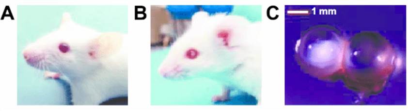 Fig.1 Cataract development in Ptch1 þ/- mice after irradiation at postnatal day 2. (A) Normal lens. (B) Cataractous lens. (C) Comparison of stereomicroscopic images of the 2 lenses. (Stefano I, et al. 2016)
Our company also offers biopsy services including:
- Slit lamp examinations, sodium fluorescein staining of the cornea.
- Direct/indirect ophthalmoscopy.
- Fundus photography (FP).
- Fundus fluorescence angiography (FFA).
- Optical coherence tomography (OCT).
- Infrared reflectance imaging (IR).
- Fundus autofluorescence (FAF).
- Intraocular pressure (IOP).
- Electroretinogram (ERG).
- Visual evoked potential (VEP).
If you are looking for the best solution for the safety evaluation of drugs for human use, please feel free to contact us.
Reference
- Stefano I, et al. (2016). "Nonlinear Radiation-Induced Cataract Using the Radiosensitive Ptch1 +/– Mouse Model." Radiation Research. 186(3): 315-321.
Related Solutions
It should be noted that our service is only used for research, not for clinical use.